- Dapatkan link
- X
- Aplikasi Lainnya
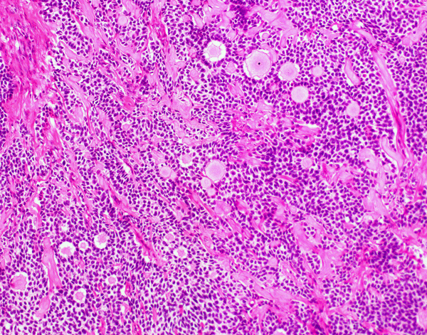
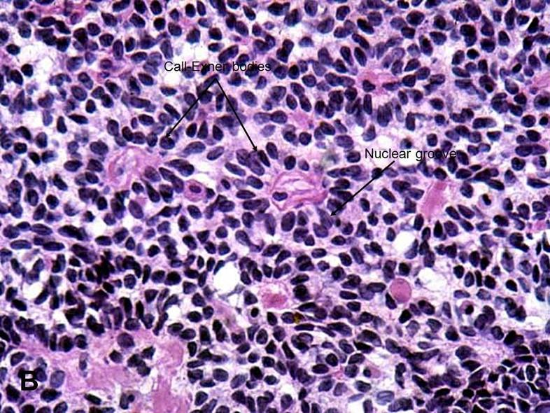
The role if any of Call-Exner bodies is not known. The granulosa cells are usually arranged haphazardly around the space.
Pathology Outlines Adult Granulosa Cell Tumor
It includes what part of the body its from and whether it was removed with surgery or a biopsy.

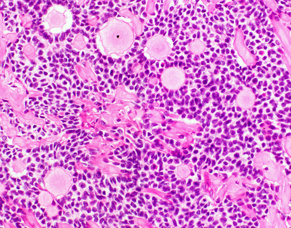
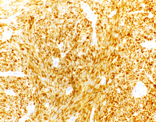
Call exner body pathology outlines. The granulosa cells are usually arranged haphazardly around the space. Call-Exner bodies are rosette-like formations with central filamentous and eosinophilic material consisting of excess basal lamina. The focus is always on excellence in.
Call-Exner bodies of ovarian follicles have been described as a ring of granulosa cells disposed radially around a central cavity filled with fluid and showing histochemical reactions similar to the liquor folliculi follicular fluid They are present in follicles of many species and in ovarian tumors. Call-Exner Bodies in granulosa cell tumor. A pathologist interprets the results of blood and pathology tests and looks for abnormalities that may point to disease such as cancer and other chronic illnesses or health risks such as pre-diabetes.
And you can also declare body parameters as optional by setting the default to None. Overview The Department of Pathology is a critical core service in the nationally-recognized Duke Health Systems and School of Medicine serving three hospitals community practices and the Durham VA Hospital. CallExner bodies giving a follicle-like appearance are small eosinophilic fluid-filled punched out spaces between granulosa cells.
Carlo Masserdotti Laboratorio DAnalisi S. There are nine specialisations in pathology. Volume 37 Issue 1 p.
Pathognomonic Call-Exner bodies which are a part of the microfollicular pattern are less common. Large round and ovoid nuclei with a longitudinal groove arranged in patterns are referred to as Call-Exner bodies microglandular pattern. The chronic lesion shows parakeratosis with lymphocytes similar to Munro microabscesses of psoriasis.
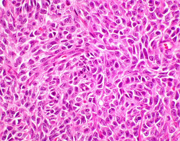
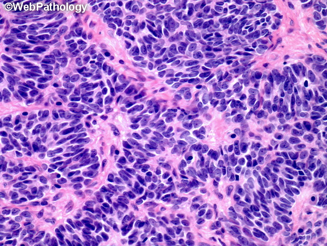
Pathology of the EndometriumPathology of the Endometrium Thomas C. In some areas neoplastic cells were in cords or columns and formed cyst-like structures. Similar cells in a stromal matrix are arranged in cords and ribbons trabecular pattern.
Cytologic detection of Call-Exner bodies in Sertoli cell tumors from 2 dogs. A variation of cells shows some cellular atypia sarcomatoid pattern. Neoplastic cells formed rosettes or Call-Exner bodies.
Purpura is often present. Wedge shaped pattern of dermal lymphocytic infiltrate. Pathology analysis The available histopathology slides of the study population were retrieved and reviewed by two experienced pathologists.
Mix Path Query and body parameters. CallExner bodies are small eosinophilic fluid-filled spaces between granulosa cells. First of course you can mix Path Query and request body parameter declarations freely and FastAPI will know what to do.
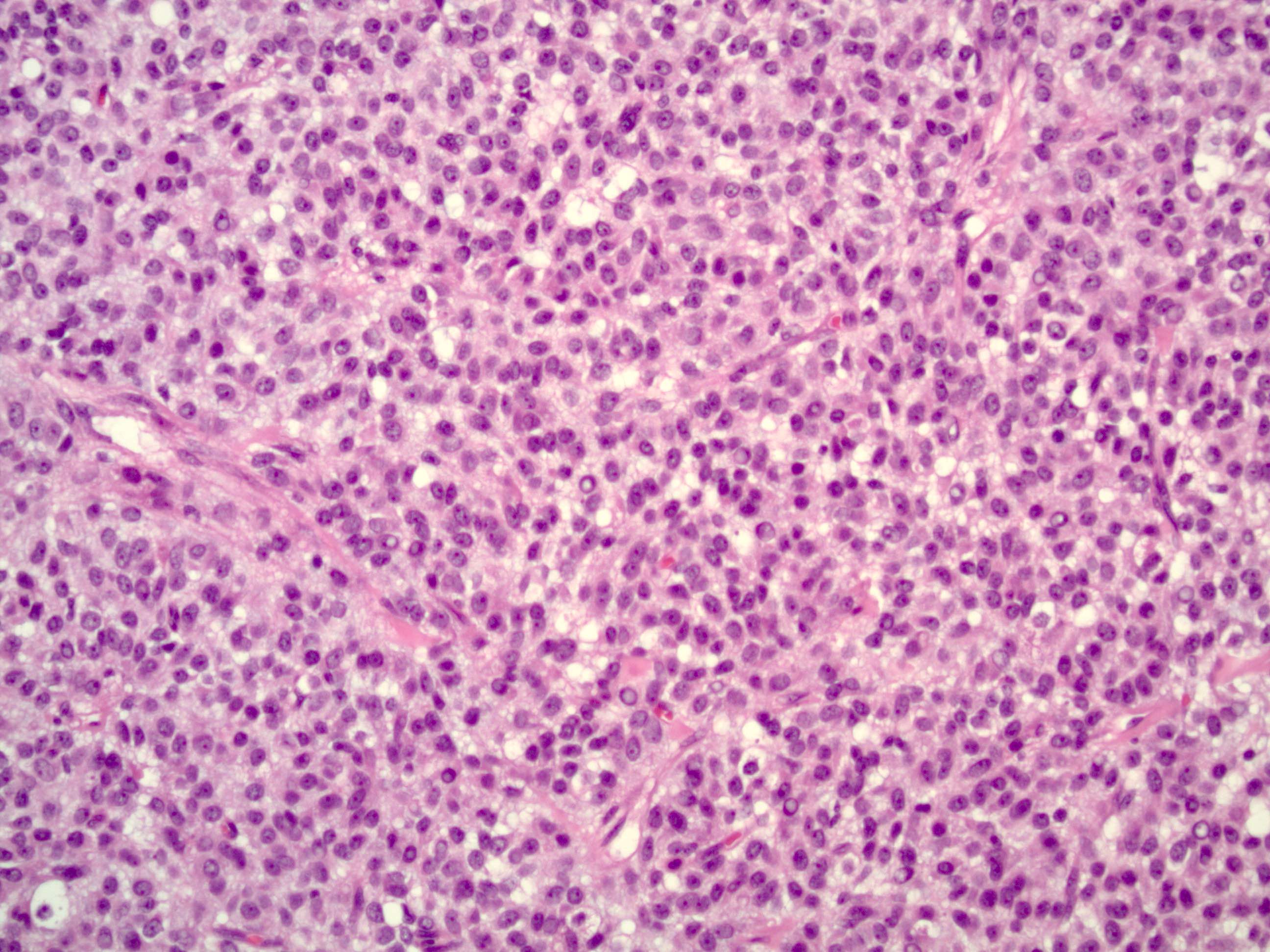
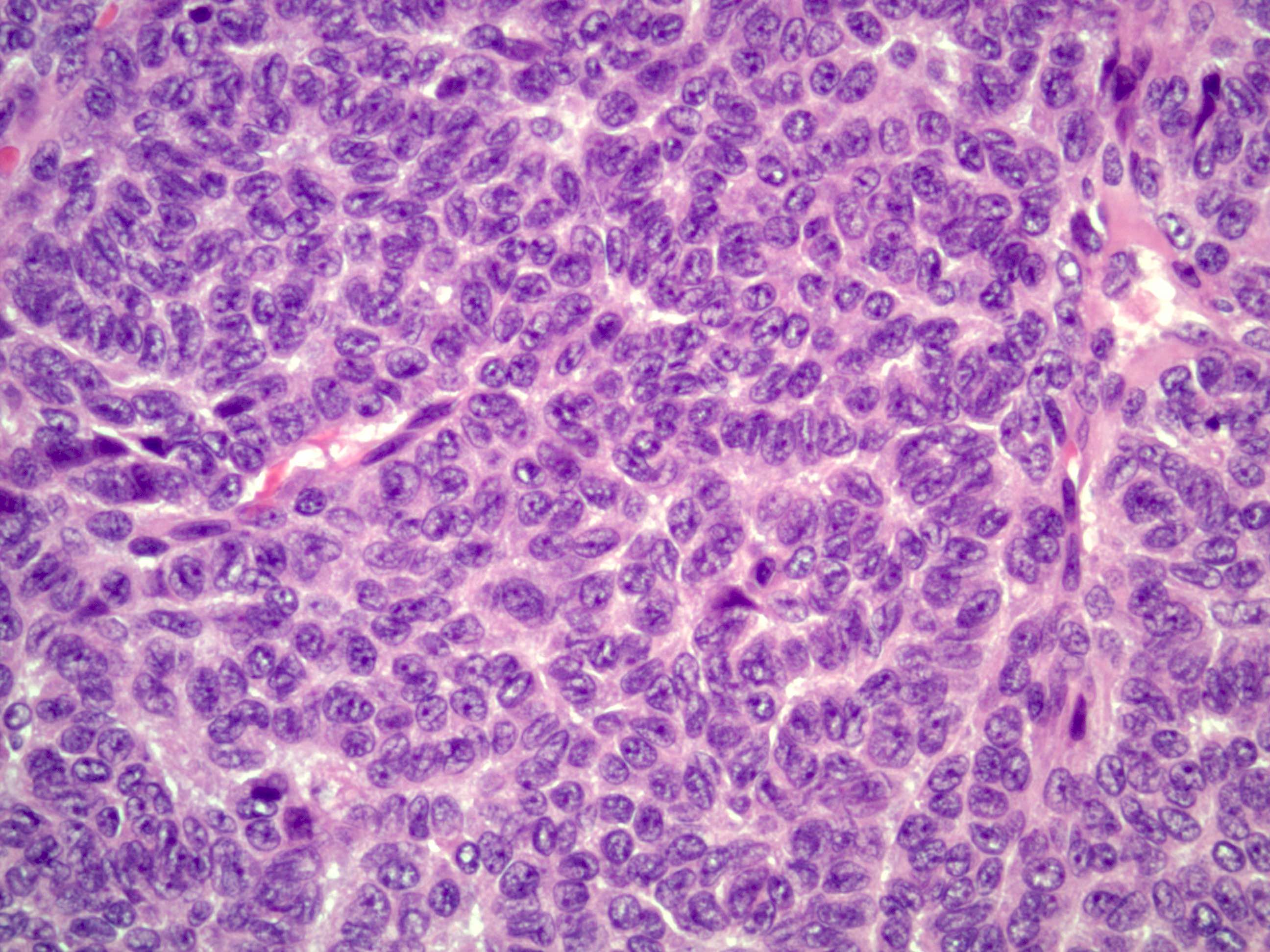
Histologically these tumors consists of monotonous islands of granulosa cells with. The Department of Pathology is composed of a large and diverse group of faculty representing all disciplines of Pathology many laboratory administrative and research staff as well as trainees and students. Morphologically the tumor cells characteristically display uniformly round to ovoid nuclei with conspicuous longitudinal nuclear grooves.
OBGyn Ultrasound 101 Mod 2. Great wolf granulosa cell tumour of ovary Medical Imaging. Veterinary Clinical Pathology.
Among these microfollicular pattern with Call-Exner bodies and coffee bean nuclei are the commonest diagnostic points2315 In the present study all four cases had microfollicular pattern and added two of the cases also showed spindlesarcomatoid and clear cell subtype. Now that we have seen how to use Path and Query lets see more advanced uses of request body declarations. Wright Columbia University New York NY Changes in the Uterus Th h t lifThoughout life.
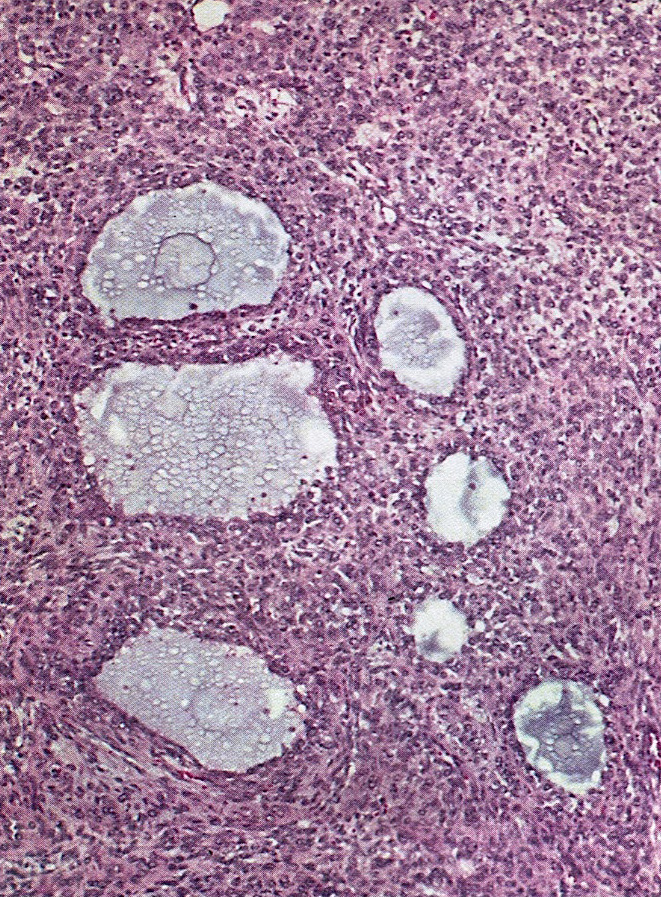
The histopathological sections of each case were assessed for the varied architectural patterns and the distinctive morphological features of GCT namely nuclear grooves and Call-Exner bodies. Microscopically these tumours are composed of small bland cuboidal to polygonal cells arranged in various patterns. An ovarian follicle with Call-Exner body-like spaces within the granulosa cell layer.
A Collection of Surgical Pathology Images. Conjuctival inclusion cysts are thin walled benign cystic lesions lined with a non-keratinizing epithelium containing serous fluid and slowly progressing cysts. Patterns included sheets of round and ovoid to spindle-shaped cells separated by thin fibrovascular stroma.
Faculty Staff Overview. Most common architectural patterns are diffuse solid insular and spindled. Encompassing surgical pathology clinical laboratories autopsy services research and bio-banking as well as educational programs Duke Pathology is an essential and integral part.
Silver stain shows reticulin fibres around clusters of cells suggestive of GCT. Call-Exner bodies Red Arrow in the image belowwhich are small follicle-like structures filled with eosinophilic material may be. Page 18 Complex Hyperplasia SldhSome glands have papillary projections into them Outlines are complex Atypical.
Management of SIL Thomas C. Vacuolar interface change and lymphocyte exocytosis less than PLEVA. The pathologist describes the tissue sample without using a microscope.
Adult Granulosa Cell Tumor. Call-Exner bodies High Quality Pathology Images of Gynecologic Ovary Sex Cord-Stromal Tumors. Pathology tests cover blood tests and tests on urine stools faeces and bodily tissues.
Parakeratosis present in well-demarcated zones. Body - Multiple Parameters. They are pathognomonic for granulosa cell tumors.
Call-Exner bodies are characteristic of GCT of the ovary with a microfollicullar pattern and they are also described in the GCT of the adult testis with the same pattern Jimenez- Quintero et.
Pathology Outlines Adult Granulosa Cell Tumor
Pathology Outlines Granulosa Cell Tumor Adult

Adult Type Granulosa Cell Tumor Humpath Com Human Pathology

Ovary Clear Cell Hyalinized Hobnail Hnf1 Histology Slides Mnemonics Medical Science
Pathology Outlines Adult Granulosa Cell Tumor
Granulosa Cell Tumor Gct American Urological Association
Pathology Outlines Granulosa Cell Tumor Adult
Pathology Outlines Granulosa Cell Tumor Juvenile
Webpathology Com A Collection Of Surgical Pathology Images
Pathology Outlines Adult Granulosa Cell Tumor

Webpathology Com A Collection Of Surgical Pathology Images Tumor Histology Slides Sinusitis

Fibrinoid Necrosis Text Pictures Small World Picture

Juvenile Granulosa Cell Tumor Tumor Cell Ovarian

Pin By Great Wolf On Granulosa Cell Tumour Of Ovary Tumor Ovaries Sarcoma
Komentar
Posting Komentar